Thursday, January 1, 2015

Tissue Repair

Tissue Repair

  • It is the process of healing after tissue injury.
  • It begins as soon as the injury begins.
  • It starts with the inflammation.

The steps are

  • Stage of Hemostasis
  • Stage of Inflammation
  • Stage of Regeneration
  • Stage of granulation tissue formation
  • Stage of remodeling

Types of wound healing 
Healing by primary intention

  • Small wound
  • Clean wound
  • Minimal contamination
  • The original tissue regenerates and heals
  • Minimal to no scarring
  • Functionally excellent
  • Occurs in tissue with labile and stable cells.

Healing by secondary intention 

  • Large, gaping wound with tissue loss
  • Infected wounds
  • Wounds with the foreign bodies inside
  • Heals with fibrosis
  • Function is impaired because of scar contracture.
  • Occurs in tissues with the permanent cells.

Stages of repair 
Stage of Hemostasis 

  • Vasoconstriction
  • Platelet aggregation
  • Clot formation

Stage of inflammation

  • Signs of inflammation
  • Neutrophils recruit in the first 24 hrs
  • Then they are replaced by macrophages after 3 days to week

Stage of fibrosis 

  • The damaged tissue is replaced by granulation tissue
  • It is mediated by different factors
  • Cytokines by macrophages like IL1 and TNF 
  • Growth factors like TGF, VEGF, PDGF, EGF
  • Granulation tissue has active fibroblasts and they are producing different ground substances
  • There is angiogenesis
  • Healthy granulation tissue looks pink in color with the serous discharge.
  • The formed collagen is collagen type III in this phase.

Stage of remodeling 
  • The scar becomes smaller due to contraction
  • It is mediated by myofibroblasts 
  • Excess tissue debris is collected by the phagocytes
  • There is regeneration of the normal tissue if possible.
  • It takes months to years.
  • Contracture of the scar causes functional anomalies.
Repair in specific organs 
Liver

  • regeneration of hepatocytes

Brain 

  • Gliosis 
  • Hydrocephalous ex vacuo

Heart

  • Fibrosis

Nerves 

  • Wallerian degeneration 
  • Nerve regeneration along the nerve sheath if the sheath is intact.

Factors effecting wound healing 
Local factors

  • Infection
  • Foreign body in the wound 
  • Wound shape and size
  • Site in the body

General factor 

  • Built
  • Nutrition
  • Co-morbid conditions
  • Rest and exercise 
  • Habits like smoking and alcohol
  • Age 

Hypertrophic scar

  • Large scar 
  • Injury site 
  • Up to 6 months 
  • Excess fibroblast 
  • Stops after sometime, no Rx
  • Type III then type I

Keloid 
  • Scar that is very large and growing 
  • May extend away from injury site
  • More than six months
  • Due to genetic predisposition
  • Rx needed, steroid or radiotherapy
  • Type III excess 


Saturday, September 20, 2014

Inflammation

Inflammation

  • It is an immediate response to injury.

Types 

  • Acute 
  • Chronic

Events 
Vascular events 

  • Increased vessel caliber and permeability 

Cellular events 

  • Recruitment of different types of inflammatory cells 

Chemical events 

  • Production of different inflammatory mediators in the site of inflammation 


Cardinal signs of inflammation

  • Redness Due to vasodilatation due to histamine  
  • Heat 
  • Swelling Due to increased vascular permeability  in respone to different chemical mediators like histamine, prostaglandins etc.
  • Pain  Due to bradykinin and p substance.
  • Loss of function 


Hemodynamic changes 

  • Initial transient vasoconstriction
  • Followed by massive vasodilation due to histamine, prostaglandins and bradykinin.

Increased vascular permeability due to

  • Vasoactive amines like histamine and serotonin
  • End product on kinin system like bradykinin
  • Cytokines like LTB4
  • Direct injury of the vascular endothelium
  • It increases the blood flow to the region and help in exudation as well as neutrophil migration.


Cellular event  

  • The most important cellular event is the neutrophil migration.
  • It is the commonest cell in the acute inflammation.
  • It is also called as PMN.
  • It has different grannules that carry the mediators of inflammation

Azurophilic grannules-

  • that contains myeloperoxidase, phospholipase A2, lysozyme and acid hydrolase.

Secondary grannules- 


  • that contain lysozyme, phospholipase A2, leucocyte alkaline phosphatase etc 

Macrophage

  • It is after 48 to 72 hrs after the injury.
  • They are large cell derived from monocyte that has lifespan for 2 to 4 months.

They have 

  • Elastase, acid hydrolases, and collagenases.
  • Produce different cytokines like interleukins and TNF, that mediate inflammation  and promotes the healing.
  • They induce the production of different growth factors like FGF, EDGF, EGF and hence help in new tissue regeneration and repair.
  • They engulf the debris that is remaining after the tissue is dead.


Neutrophils migration and defence 
Steps 

  • Vasodilation and rolling
  • Marginalisation
  • Adhesion
  • Diapedesis 
  • Chemotaxis 
  • Phagocytosis and degranulation 
  • Intracellular killing 


  • Adhesion is due to the surface molecules on the endothelium and the neutrophils like selectin and integrin respectively.
  • These molecules are expressed due to other different chemicals like IL1, TNF etc.

Defect in adhesion occurs in 

  • DM
  • Steroid therapy
  • Uremia
  • Alcohol intoxication 
  • Leukocyte adhesion defect (autosomal recessive cond.)

Chemotaxis

  • It is the attraction of the inflammatory cells at the site of the tissue injury by the use of chemicals.

Types:

  • Bacteria products like N-formyl methionine
  • Inflammatory mediators like 
  • LTB4- product of arachidonic acid
  • Complement proteins like C3a, C5a
  • Chemokines like IL1 produced by macrophages. 


Leucocyte migration 


Phagocytosis 

  • It is the process of engulf the bacteria by neutrophils 

How does neutrophil recognizes the bacteria? (opsonins)

  • Coated by antibody
  • Coated by complement C3b
  • Plasma proteins like collectins
  • The engulfed bacteria or product is called phagosome and when it combines with the lysosomes, it becomes phagolysosomes.


Intracellular killing 
Types
Oxygen dependent- 

  • Using superoxide radicals with the use of  NADPH oxidase and producing superoxide and superchloride ions.

Oxygen independent killing

  • Using lysozyme, lactoferrin, acid hydrolases which make pore in the bacterial cell wall and then causing the lysis of the bacteria. 
  • NADPH oxidase deficiency leads to the disease called as chronic granulomatous disease.


Chemical mediators of inflammation 


Mediators of inflammation 


Complement system 

  • They are a group of proteins that are produced by the liver

They have different functions,

  • C3a, C5a- chemotaxis and anaphylotoxins 
  • C3b- works as opsonin  so it is target for phagocytosis and intra or extracellular killing
  • C5b-C9- they form membrane attack complex.


TNF and IL 

  • They are produced by activated macrophages 

They have the following function

  • Fever 
  • Apoptosis
  • Decrease apetite and cachexia
  • Insomnia 
  • Stimulate the production of the different growth factors like EDGF, FGF, TGF etc.
  • IL 8 is a chemoattractant.


Outcomes of inflammation 

  • Complete resolution with regeneration 
  • Complete recovery with scarring 
  • Chronic inflammation
  • Abscess formation


Chronic inflammation 
Causes 

  • Acute inflammation
  • Persistent infections like TB
  • Autoimmune disease
  • Foreign body inside body
  • Malignancy 


Key players in the chronic inflammation 
Macrophage 

  • Modified macrophage is called epithelioid cell.
  • Fused multiple macrophages makes giant cell.

Lymphocytes 

  • Types, B cell and T cell.
  • B cell produce antibodies and T cells help B cells and also help cell mediated immunity.

Eosinophills and basophills 

  • mainly in the allergic conditions and worm infestations.
  • Eosinophils has a protein in the cytoplasm called as major basic protein, that is toxic to the parasites.


Granulomatous inflammation 

  • It is special type of inflammation in which the infecting organisms living or dead are surrounded by macrophages(epithelioid cell and giant cell) and lymphocytes Th1, as well as plasma cells.

Key mediators are 

  • IFN gamma
  • TNF alfa
  • IL 2


  • All are produced by activated T cells and macrophages.
  • They convert the macrophages to epithelioid cells and giant cells.
  • Common example is TB. If someone is taking TNF inhibitor like in RA like Infliximab and Etarnacept , they should do PPD test, otherwise they reactivate the latent TB.


Histologic patterns/types of inflammation 
Exudative inflammation

  • Vascular permeability increases causing fluid and protein leak out of the blood vessel.
  • Eg. Pneumonia, meningitis

Necrotizing inflmmation

  • When inflammation causes an extensive tissue damage.
  • E.g. Forunier gangrene caused by gram negative bacteria.

Granulomatous inflammation


  • Slow growing organism like TB, fungi etc


Interstitial inflammation 
  • Like in the glomerulonephritis.
  • Recruitment of the inflammatory cells in the interstitium.
Cytopathic inflammation
  • When the infected cell in the tissue is altered.
  • Mainly in viral infection like CMV inclusion bodies, Negri bodies in the rabies.
No inflammation despite infection
  • Immunocompromised state like AIDS, steroids therapy, organ transplant receiver etc.


Sunday, June 22, 2014

Cell Injury and Adaptation

Cell injury 
Causes are 

  • Hypoxia 
  • Infections
  • Immunologic reactions
  • Congenital disorders 
  • Chemical injury
  • Physical injury
  • Nutritional imbalance 


Hypoxia

  • Deficiency of oxygen due to redn in blood flow.

Types:

  • Anoxic or hypoxic
  • Anemic 
  • Cardiopulmonary failure
  • Histotoxic 


Infections

  • Bacterial 
  • Viral 
  • Protozoal 
  • Fungal
  • Proteins like prions induced disease


Immunological reactions 

  • Also called hypersensitivity reaction.
  • They are protective to our body, but when excess they can have collateral tissue damage.
  • Autoimmune diseases are targetted to the body cells causing their destruction.

Types:

  • Type I
  • Type II
  • Type III
  • Type IV 


Congenital disorders 
Disease due to trisomy

  • Down’s syndrome
  • Patau synrome
  • Edward syndrome

Disorders of sex chromosome 

  • Klinefeter’s syndrome
  • Turner’s syndrome

Autosomal recessive 

  • Osteogenesis imperfecta
  • Cystic fibrosis
  • Alkaptonuria
  • Phnylketonuria
  • Glycogen and lysosome storage disease


Autosomal dominant

  • Familial hypercholesterolemia
  • Marfan syndrome
  • Ehler Danlos syndrome
  • Neurofibromatosis

X linked recessive

  • Lysch nyhan syndrome
  • Chronic granulomatous disease

X linked dominant

  • Fragile X syndrome
  • Huntingtons disease


Physical agent

  • Heat
  • Cold
  • Radiation
  • Burn
  • Crush
  • Gunshot 
  • Pressure changes


Chemicals 

  • Acid
  • Alkali
  • Cyanide
  • Poisons like arsenic, lead, mercury
  • Alcohol
  • Cigarette smoking
  • IV drug abuse 


Nutrition
PEM 

  • Anorexia nervosa
  • Kwarshiorker
  • Marasmus

Vitamin deficiency

  • Scurvy, beri beri, pellagra
  • Anemia
  • Subacute combined degeneration (vit B12)

Nutrition excess


  • Obesity and related disorders

Tissue response 


Cellular changes during cell injury

Reversible cell injury 

  • ATP depletion
  • Glycolysis and acidosis
  • Na/H2O influx and cell swelling
  • Ribosomal detachment
  • Plasma membrane blebs and myelin figures are seen


  • If oxygen is available and the other noxious stimuli are removed the cell can recover. If not it will go into irreversible stage.

Irreversible cell injury hallmarks 

  • Severe membrane damage
  • Marked mitochondrial dysfunction
  • Lysosomal rupture and cell autodigestion
  • Nuclear changes 
  • Condensation/ pyknosis 
  • Fragmentation/ karyorrhexis
  • Disappearance / karyolysis 


Finally it will lead to the cellular death and inflammation.

Tissue death/necrosis 
Morphologic types 

  • Coagulative necrosis- due to ischemia, cells are dead but cellular architecture is preserved. Eg in solid organs like liver and kidney.
  • Liquefactive necrosis- necrosed cells are partially digested by lysosomal enzymes, and hence the cellular architecture is lost revealing cheesy material. Eg in brain , abscess and spleen.
  • Casseous necrosis- combination of these two types. Seen in granulomas like TB.
  • Fat necrosis- in fatty tissue due to the release of lipase.
  • Fibrinoid necrosis- with the deposition of non specific protein. That results from acute immunologic injury. Eg Aschoff bodies in RHD,  Rheumatoid nodule, hypertensive vasculitis etc.

Gangrene-

  1. it is the gross necrosis of a large tissue.

Dry gangrene- coagulative necrosis

Wet gangrene- liquefactive necrosis

Apoptosis

  • Programmed cell death without inflammation.

Morphology:

  • Cell shrink
  • Nuclear condensation 
  • Nuclear fragmentation
  • Cellular fragmentation into apoptotic bodies 
  • Phagocytosis

Pathway 

  • Intrinsic
  • Extrinsic 


Examples of apoptosis 
Physiological 

  • Organogenesis 
  • Menstruation
  • Lymphocytes in the thymus 

Pathological 

  • Viral hepatitis- councilman bodies
  • Cystic fibrosis- pancreas atrophy


Serum markers 

  • When the cells die, their enzyme comes out and level can be detected in the blood.
  • This gives an important clue for the diagnosis of the disease.

Common markers are

  • AST and ALT
  • CKMB
  • Amylase and lipase
  • LDH
  • Myoglobin 


Tissue adaptation to injury 
Atrophy 

  • Decrease in organ size due to decrease in the number and the size of each cell.

Cause of atrophy are

  • Disuse due to immobilisation
  • Denervation atrophy
  • Lack of hormone stimulus
  • Malnutrition 
  • Aging

Picture:

  • Decreased cellular components
  • Autophagolysosomes 


Hypertrophy

  • Increase it the organ size due to the increase of the cell size.

Causes:

  • Increased Mechanical demand like weight lifters or in hypertension (cardiac hypertrophy)
  • Increased hormone stimulation e.g. gravid uterus , lactating breast.

It occurs in response to

  • Growth factors
  • Cytokines
  • They lead to increased expression of the concerned genes and then causing increased protein synthesis.


Hyperplasia

  • Increase in the organ size because of the increase in the number of cells.

Cell types:

  • Labile cells
  • Stable cells
  • Permanent cells 
  • Only stable and permanent cells can undergo hyperplasia.

Eg:

  • After hepatectomy, the liver gains full size. Breast development in puberty etc.
  • The stimulus are similar the hyperplasia.


Metaplasia

  • The reversible change of one cell type to another.
  • It is in response to chronic irritation.

Examples are

  • Squamos metaplasia of the bronchail epithelium in the smokers
  • Barret’s  esophagus
  • The new type of epithelium come from the reserve cells that lie in the basal layer.
  • It is not premalignant, but chances of malignancy is as high as 10-15 times than the normal.


Dysplasia

  • The loss of cell differentiation is called dysplasia.
  • It is the hallmark of malignancy.
  • It is characterized by changes in cell shape, size and differentiation.

Examples 

  • Oral leukoplakia
  • Cervical dysplasia
  • Solar keratosis 


Wednesday, June 11, 2014

Pathophysiology of Cell


Pathology 

  • Patho- suffering 
  • Logus- study 

Physiology 

  • Physio- function 
  • Logus- study 

Pathophysiology

  • study of sequence of events from normal function to suffering.

Parts of cell 
  • Membranes 
  • Nucleus 
  • DNA
  • Euchromatin
  • Heterochromatin
  • Histone proteins   
  • Nucleolus 
  • Cytoplasm 
  • Matrix
  • Organelles
  • Ribosomes
  • Endoplasmic reticulum
  • Mitochondria
  • Golgi apparatus
  • Lysosomes
  • Peroxisome
  • Cytoskeletal molecules 


DNA 

  • Contains all genetic info.
  • Arranged in fibers with the histone proteins, in 30 nm or 10 nm solenoid fibers.
  • Condensed DNA at the time of cell division is called chromatin.

Made of

  • Sugar- pentose sugar
  • Nitrogen base – two types 
  • Phosphate backbone 

Take part in two events

  • Replication- structural importance 
  • Transcription- functional importance

Any unwanted modification in the structure is the cause of genetic diseases and cancer.

RNA 

  • Transcription product of DNA
  • Carries message from the nucleus into the cytoplasm.
  • The genetic info is read by ribosomes and that is decoded into the proteins.

Made of

  • Deoxyribose sugar
  • Nitrogen bases 
  • Phosphate backbone

Three types:

  • Ribosomal RNA- makes up the ribosome
  • Messanger RNA- takes the info from the nucleus
  • Transfer RNA – carries needed amino acids from the cyto into the nucleus.


Cytoplasm
Ribosomes:

  • Small particles in the cytoplasm made of rRNA and protein 
  • Either free or membrane bound with RER
  • Human ribosomes are 80’s ribosome (bacterial are 70’s)
  • Have two subunits, 60’s and 40’s
  • Polysomes 

Function:

  • Protein synthesis by traslation 
  • Ribosomal detachment occurs at the time of cell injury is an important event, which limits the protein synthesis in the cell that leads to the irreversible injury and cell death.


Endoplasmic reticulum 
They are sac like structures attached to the outer nuclear membrane.
Types:

  • Smooth- without endoplasmic reticulum
  • Rough- with endoplasmic reticulum.

Functions:

  • RER systhesizes protein for secretion, receptor proteins and lysosomes.

SER 

  • Lipid synthesis and metabolism
  • Drugs detoxification
  • Glucose systhesis by glycogenolysis and gluconeogenesis 
  • Release calcium in skeletal muscle 


Lysosome
They are simply the packets of the enzymes like acid hydrolases, nuclease, glycosidase, lipase protease etc in the cytoplasm.
Types:

  • Primary lysosome
  • Secondary lysosome 

Function:

  • Intracellular digestion of the cell debris and endocytosed foreign body.

Pathological concerns:

  • If some enzymes are deficient, then lysosomal storage disease like, Tay Sach’s disease( deficiency of hexaminidase, leading to accumulation of galglioside GM2 in the brain cells and mental retardation), Gaucher’s disease (deficiency of glucocerebidase),  Niemen Pick’s disease ( deficiency of sphingomyelinase) etc.
  • Membrane rupture and release of the lysosomal enzymes lead to the intracellular digestion and plays an important role in the cell death and apoptosis.


Mitochondria 
It is double membrane structure, with the inner membranes are folded called as cristae, to increase the surface area.
It is the organelle that has its own DNA.
This DNA is only derieved from the mother, rest all are from the both.
So, any mitochondrial disease like Leber’s optic neuropathy, male infertility due to mitochondrial dysfunction is only inherited from the mother.
The part inside the inner membrane is called matrix, that has the dehydrogenase enzymes for the oxidative phosphorylation of NADH and FADH2 and generation of ATP.
So mitochondria is called as the powerhouse of the cell.


Mitochondria and relations to the pathology are

  • Hereditary mitochondrial diseases like Leber’s optic neuropathy
  • There is a protein called cyrochrome  c, that is an iron containing pigment uses in the oxidative phosphorylation. If this protein is released at the time of cellular injury, it directs the cell towards apoptosis.
  • Mitochondrial dysfunction in the cell injury is a very critical step in the irreversible cell damage. Without mitochindria, the ATP’s out of a glucose molecule is just 2, with the production of lactic acid. And poor supply of ATP and acidosis will lead to the cell death very quickly.


Peroxisome 
They are a single membrane bound vesicles.
They contain the enzymes like peroxidase and catalase, that are related with the production and destruction of superoxide ions.
Their function are

  • Provide free radicals during extracellular killing of foreign pathogens.
  • Beta oxidation of long chain fatty acids
  • Bile acid systhesis.


Pathology concerns 

  • There are some genetic disorders like Zelweger syndrome, Refsum disease, infantile adrenoleukodystrophy  in which the peroxisomes are deficient and hence the long chain and branched fatty acids as well as the precursors of the bile acids are accumulated in the cell and causing cell dysfunction.
  • The affected individual will have scanty myelin in CNS, progressive deafness and blindness, and MR, repeated infections and the death in ZS is as early as before 1st birthday.


Cytoskeleton 
Types:
1. microfillment- smallest one 7nm

  • Actin
  • They in conjunction with mysoin make the contractile unit
  • They help in diakinesis in cell division.
  • They form the core of the microvilli.

2. intermediate fillament

  • Type i- Keratin- skin
  • Type ii- Desmin- muscles, Vimentin in fibroblasts, myofibroblasts, and endothelium as well as vascular smooth muscles
  • Type iii. Neurofillaments 
  • Type iv- Lamins in the nuclear membrane.

3. micritubules- Largest 25 nm hollow tubes 

  • Are attached with the ATPase motor molecule, that cleaves the ATP and provides the energy.
  • The help in retrograde (Dyenin) and anterograde (Kinesis) transport. Speed is 200-400mm/d anterograde transport, and 4-200mm/d retrograde transport.


Pathology significance 
  • Many virus like HSV and rabies as well as polio virus are stored in the ganglia, where they reach with the help of the retrograde transport.
  • Cancer cells have augmented synthesis of these cytoskeletal molecules and hence fast cell division. So these are the targets of many anticancer drugs.
  • In many diseases, there is deposition of these fibers. Like in cirrhosis, there is deposition of intermediate fillaments like keratin (mallory bodies), and Alzheimer's disease, there is deposition of the neurofibrillary tangles in the certain memory areas like hippocampus.
Cell adhesion molecules 
Calcium dependent

  • Cadherin 
  • Selectin

Calcium independent 

  • Integrins 

Function

  • Adheres the cells between each other and the basal lamina 
  • Leucocyte migration into the tissue, integrin in the neutrophils binds to the selectin in the endothelial cell and gets in to the tissue.

Disease:
Antibodies to the desmosomes will cause pemphiguous vulgaris, and hemidesmosome will cause bullous pemphigoid.
Disruption of the cadherin molecule in the BM is the first step in the mets of the cancer.

Tissue
Group of similar cells for similar function.
Types:

  • Epithelial tissue- origin ectoderm and endoderm
  • Connective tissue- mesodermal in origin
  • Nervous tissue- neuroectoderm and neural crest in origin
  • Muscular tissue- origin mesoderm.

Pathological importance:

  • Some disease involve single tissue and Some involve multiple
  • Some  disease are primary to that tissue like muscular dystrophy, some are secondary or reactive like RHD.


Epithelial 
On the basis of striations 

  • Simple 
  • Striated

On the basis of cell structure

  • Squamos 
  • Columnar
  • Cuboidal
  • Transitional 

On the basis of cillia

  • Cilliated 
  • Non cilliated 


Muscular tissue 
It is the contractile tissue
It has actin and myosin, that glide on each other and hence they are contractile.
Types:

  • Skeletal muscle
  • Cardiac muscle
  • Smooth muscle.


Connective tissue 
They are the supporting tissue.
Types:  
  • Dense - bone, cartilage, dentine, enamel
  • loose- dermis, adipose tissue etc
  • Specialized connective tissue like blood, bone marrow, cartilage, adipose tissue.

Constituent 

  • Cells- fibroblast, myofibroblast, chondroblast, osteoblast, adepocytes, and mast cells.  Histiocytes or Langerhans cells are blood borne macrophages that live in tissue.
  • Extracellular matrix made of fibrillar protein (collagen, elastin, fibrillin, fibronectin), gel matrix made of preteoglycans and glycoproteins.
  • Many  connective tissue diseases are targetted to them, like SLE, RA etc.

Main disease related with the bone 

  • Rickets and osteomalacia- vitamin D deficiency
  • Paget’s disease 
  • Osteoporosis 


Types of collagen 

  • Type I- in bones, cartilage, skin, sclera etc
  • Disease is Osteogenesis imperfecta (mutation in gene coding type I cartilage)
  • Type II- cartilage
  • Type III- reticular fibers in the blood vessels.
  • First formed collagen in the wound healing, that is later replaced by type I.
  • Type IV is found only in the basal lamina, in the lamina propria.

Diseases are:

  • Scurvy- inadequate hydroxylation of the preprocollagen
  • Ehler Danlos syndrome
  • Marfan syndrome- deffect in fibrillin gene.

Nervous tissue 
Types :

  • excitable tissue ie neurones 
  • Non excitable tissue like supporting cells.

Neurones:

  • Unipolar
  • Bipolar
  • Multipolar

Glial cells:

  • Astrocytes
  • Oligodendrocytes
  • Ependymal cells
  • Microglia and 
  • Schwaan cells 

CNS does not have fibroblasts. (clinical correlation is so brain tissue has no fibrosis after injury, but gliosis or hydrocephalous ex vacuo)


Disease concern 
  • Gullian barre syndrome and multiple sclerosis- relation with myelin 
  • Alzheimer's disease nerve degeneration with deposition of non specific protein material in the CNS
  • Huntington’s disease degeneration of neurones in the caudate nucleus
  • Parkinsonism neuronal degeneration in the nigrostiatal pathway
Types of Epithelium

Pathologic concerns with the blood 

Inflammation:

  • 1st responder- neutrophil
  • 2nd responder- macrophage, lymphocytes

Bone marrow malignancy
Immunity and related disorders
Anemia 

Tuesday, May 13, 2014

Pelvis And Perineum

Pelvis
Composition:

  • formed by paired hip bones, sacrum, coccyx, and their articulations

Two portions 

  • Greater pelvis
  • Lesser pelvis
  • Terminal line ( pelvic inlet): formed by promontory of sacrum, arcuate line, pectin of pubis, pubic tubercle, upper border of pubic symphysis
  • Pelvic outlet: formed by tip of coccyx, sacrotuberous ligament, ischial tuberosity, ramus of ischium, inferior ramus of pubic symphysis

Muscles of pelvic wall
  • Piriform m.
  • Obturator internus m.
Muscles of floor of pelvis
Levator ani
  • Levateo prostate (pubovaginalis)
  • Puborectalis
  • Pubococcygeus  and iliococcygeus
  • Coccygeus
Pelvic  diaphragm
  • Superior fascia of pelvic  diaphragm
  • Levator ani
  • Coccygeus
  • Inferior fascia of pelvic  diaphragm
Pelvic fascia 
Parietal pelvic fascia 

  • A continuation of the transverse fascia into the pelvis. It coves the piriformis and obturator internus 
  • Attaches to the arcuate line of the pubis and ilium, thickens over the obturator internus to form the arcus tendineus, the origin of portions of the levator ani muscle
  • At the tendinous arch of levator ani it splits to cove both superior and inferior surfaces of the levator ani as superior and inferior fascia of pelvic diaphragm 

Visceral pelvic fascia 
  • Lies between the peritoneum and the pelvic viscera
  • It is a continuation of the extra peritoneal connective tissue
  • Ensheathes retroperitoneal viscera and forms septa between retroperitoneal organs
  • Rectovesical septum        
  • Rectovaginal septum         
Retropubic space        
  • Lies between pubic symphysis and urinary bladder
Pararectal space 
  • Lies around the rectus 
Retrorectal space

Common iliac artery

  • Continuation of abdominal aorta at level of L4 vertebra 
  • Terminates in front of sacroiliac joint by dividing into internal and external iliac arteries 


Internal iliac artery 

  • It is a terminal small branch of the common iliac artery.
  • It is about 4 cm long.
  • In the foetus, this artery is the origin of the umbilical arteries to the placenta.
  • It begins in front of the sacroiliac joint, between L5 and sacrum, medial to psoas muscle, and runs backwards and ends near greater sciatic notch by dividing in to two branches, the anterior and the posterior division.

The relations are:

  • Anterior- ureter
  • Posterior- internal iliac vein
  • Lateral- external iliac vein and the obturator nerve
  • Medial- peritoneum and the tributaries of the internal iliac vein.

Anterior division

  • Superior vesicle artery
  • Obturator artery
  • Middle rectal artery
  • Inferior vesicle artery
  • Inferior gluteal artery
  • Internal pudendal artery
  • Vaginal artery
  • Uterine artery


Posterior division 
  • Illiolumbar artery 
  • Lateral sacral artery
  • Superior gluteal artery
Uterine Artery
  • about 2cm from neck of uterus it crosses above and in front of ureter
Internal pudendal artery
  • Perineal artery
  • Anal  artery
  • Dorsal  artery of penis (clitoris) 
Veins of pelvis
Internal iliac vein

  • Parietal tributaries: accompany with arteries
  • Visceral tributaries
  •                  →superior rectal vein→inferior mesenteric v.
  • ①Rectal venous plexus    →inferior rectal vein→internal iliac v.
  •    →anal vein→internal pudendal v.   
  • ②Vesical venous plexus →vesical v.
  • ③Uterine venous plexus →uterine v. 
  • External iliac– accompany the artery
  • Common iliac vein– formed by union of internal and external iliac veins in front of sacroiliac joint, end upon L4~L5 by uniting each other to form inferior vena cava


The lymphatic drainage of pelvis
Internal iliac lymph node

  • Surround internal iliac vessels
  • Receive afferents from pelvic viscera, perineum, buttock and back of thigh

External iliac lymph nodes 

  • Lie along external iliac artery
  • Receive afferents from lower limb and some parts of pelvic viscera
  • Sacral lymph node

Common iliac lymph node 

  • Lie along common iliac artery
  • Receive afferents from all the above nodes
  • Efferents pass to lumbar lymph node 


Sacral plexus
Formation:

  • formed by anterior rami of L4 and L5 spinal nerves (the lumbrosacral trunk) and anterior rami of sacral and coccygeal nerves


Position:

  • lies in pelvic cavity, anterior to sacrum and piriformis 

Branches
  • Superior gluteal 
  • Inferior gluteal 
  • Pudendal
  • Posterior femoral cutaneou 
  • Common Sciatic 
  • Autonomic plexuses of plevic
  • Hypogastric plexus 

  • Superior hypogastric plexus : lies in front of L5 between common iliac ateries
  • Inferior hypogastric plexus (pelvic plexus): lie on each side of rectum

Sacral sympathetic trunk

  • Is continuos above with the abdominal part
  • Has 4or 5 ganglion 


Relationships of rectus
Anteriorly

  • In male 
  • Fundus of bladder
  • Seminal vesicle
  • Prostate 
  • Ampulla ductus deferentis
  • In female 
  • neck of uterus
  • Vagina 

Posteriorly

  • Sacrum and coccyx
  • Piriformis
  • Median sacral vessels
  • Anterior branches of sacral and coccygeal nerves
  • Sacral sympathetic trunk

Laterally 

  • Pelvic  plexus
  • Superior and inferior rectal vessels
  • Levator ani  

Vessels and lymphatics of rectum
Arteries 

  • Superior rectal a.
  • Inferior rectal a.
  • Median sacral a.
  • Lymphatic drainge Lymphatics follow arterial blood supply to following nodes:
  • Superior rectal ln.
  • Inferior mesenteric ln. 
  • Pararectal ln.
  • Internal iliac ln.
  • Sacral ln.

Perineum
General features

  • Region of below pelvic diaphragm
  • A diamond-shape space whose boundaries are those of the pelvic outlet
  • Lower border of symphysis pubis
  • Rami of pubis and ischium
  • Ischial tuberosities
  • Sacrotuberous ligament 
  • The coccyx


Two triangles
 An imaginary line drawn between the two ischial tuberosities divides perineum into anterior and posterior triangles

  • Urogenital region (anterior)-differs in male and female
  • It has urogenital diaphram, which is made by,
  • Muscles- sphincter urethrae and deep transverse perinei
  • Fascia- superficial and deep urogenital fascia 
  • Anal region (posterior)-similar in both sexes 

Branches
  • Superior gluteal 
  • Inferior gluteal 
  • Pudendal
  • Posterior femoral cutaneou 
  • Sciatic Common
Urogenital region 
Superficial fascia has two layers

  • The superficial or fatty layer
  • The deep or membranous layer (superficial fascia of perineum or Colles fascia) 

 Anteriorly- it is continuous with:

  • Dartos of the scrotum
  • Fascia of the penis
  • Membranous layer of superficial fascia of the abdominal wall known as the fascia of Scarpa

Deep fascia has two layers

  • Superior fascia of urogenital diaphragm
  • Inferior fascia of urogenital diaphragm


Superficial perineal pouch 
Boundaries

  • Lies between inferior fascia of urogenital diaphragm and superficial fascia of perineum
  • Space open anteriorly (In rupture of cavernous part of urethra, urine can extravasate from scrotum upward in front of symphysis pubis into anterior abdominal wall deep to membranous fascia of Scarpa)

Contents

  • Posterior part-superficial transverse perineal muscle
  • Lateral part-crus penis (male), crus of clitoris (female) and ischiocavernousus covering them
  • Central part-bulb of urethra (male), bulb of vestibule (female) and bulbocavernousus covering them
  • Branches of pudendal nerves and internal pudendal vessels


Deep perineal space
Lies between superior and inferior fascia of urogenital diaphragm
Contents 

  • Deep transverse perineal muscle
  • Bulbourethral gland (male)
  • Sphincter of urethra (male),urethrovaginal sphincter
  • Ateries, veins and nerves


Urogenital diaphragm 
Triangular in shape
Attached laterally to ischiopubic rami and ischial tuberosities

Formed by sphincter of urethra, deep transverse perineal muscle, superior and inferior fascia of urogenital diaphragm
perineal body

  • Wedge-shape fibromuscular mass
  • In female, between anal canal and lower end of vagina, 
  • In male, between anal canal and root of penis
  • It is larger in the female than in the male and five support to the posterior wall of the vagina


Origin or insertion of several small muscles and insertion of part of palvic diaphragm
These  muscles are: 
  • Sphincter ani externus 
  • Levator ani 
  • Superficial transverse muscle perineum 
  • Deep transverse muscles perineum 
  • Bulbocavernousus  
  • Sphincter of urethra (male) or urethrovaginal sphincter (female)
Pelvic organs

  • Male reproductive organs
  • Female reproductive organs
  • GI organs like rectum and anal canal
  • Urinary system organs like distal ureter, bladder and the urethra.


 Urinary structures 
Ureter:

  • They are narrow paired tubes that convey the urine to the bladder.
  • 25 cm long and 3mm in diameter.
  • Upper half is in the abdomen and the lower half in the pelvis.

Course:

  • Begins from the renal pelvis.
  • Passes downwards and slightly medial to the psoas muscle.
  • Crosses the external iliac artery near its origin from the common iliac artery to reach the pelvis.
  • Runs along the greater sciatic notch, medially and posterior.
  • Then comes anterior and medially to reach the base of the bladder. 


Constrictions:
  • At pelviureteral junction
  • At when it passes into the pelvis, crossing the internal iliac artery
  • At its insertion into the bladder, called as vesicoureteral junction.
Blood supply:
  • Upper part- renal artery
  • Middle part- branches of the aorta
  • Inferior part- branches from the vesicular, middle rectal and uterine vessels.
Nerve-
  • smpathetic and parasympathetic from the T10-L1 and S2-4 respectively.
It has an important relation with the uterine artery in its forward course, it passes beneath the uterine artery in the extraperitoneal sheath between the medial and the lower part of the broad ligament. Here it can be ligated or cut during the hysterectomy.
Urinary bladder 

  • It is the site of temporary storage of urine.
  • The size, location and shape depends on the amount of the urine that the bladder contains.
  • It is in the pelvis when empty, but comes to the abdomen when its full.

When empty, it is pear shaped with the following parts:

  • Apex forward
  • Base backwards
  • Neck below attached with the urethra and
  • Three surfaces- superior, right and left inferolateral

Relations:

  • Anterior: apex is connected to the umbilical by median umbilical ligament (degenerated urachus).

Base/posterior- 

  • cervix and vagina in females
  • males- rectum superiorly by rectovesical pouch, and inferiorly fascia of denonvillers with the seminal vesicles and the vas deferens.

Neck-

  • Males- related to the prostate gland.
  • Female- it lies in the pelvic fascia.

Superior-

  • its covered by the peritoneum.


Urethra
It is the organ for the external drainage of the urine.
It has dual function in males:

  • Ejaculation
  • Urine voiding.
  • But is has only the urinary function in the female.
  • The length is 15-20 cm in the males, and it is only 4 cm in females.
  • Blood is supplied by the branches from the prostatic vessels and the penile vessels.

Parts:

  • Prostatic part (3cm)- beneath the prostate gland
  • Membranous part(2cm)- inside the pelvic diaphragm
  • Spongy or penile part(15 cm)- inside the corpus spongiosum in the penis, ventral surface.
  • The prostatic urethra is only the homologus part of the female urethra.


The opening of the urethra is guarded by two sets of sphincters:
Internal sphincters
  • they are just the condensation of the muscles of the bladder in circular fashion around the neck of the bladder.
  • They are supplied by the autonomic fibers.
  • They are functional in man, and less in woman.
  • Their function is the control of the retrograde ejaculation, no the urine continence.
External sphincter:
  • Located in the urogenital diaphragm.
  • Has circular muscles, called as sphincter urethrae.
  • It is supplied by pudendal nerve.
  • It is under voluntary control.
  • They are developed in both males and the females.
  • Function is the urinary continence. 
Lymphatic drainage: 
  • from the prostatic and membranous part is to the internal iliac nodes, and
  •  from the penile part is to the deep inguinal nodes.
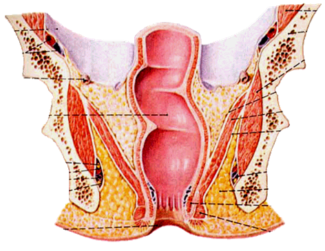
Rectum

  • It lies between the sigmoid colon and the anal canal.
  • When it is full, person will have urge to defecate.

Location:

  • In the true pelvis, in front of the S3-S5.
  • Length is 12 cm and is 4 cm wide.

It has two curves:

  • anteroposterior- first anterior, then posterior.
  • Lateral- first convex to right, then left and then again right.


Relations
Peritoneum:

  • Upper third- covered by peritoneum from the sides and the front
  • Middle 3rd- only front
  • Distal 3rd- no peritoneum.

Anterior:

  • Males- rectovesical pouch with intestines, urinary bladder, seminal vesicle and prostate from up to down.
  • Females- rectouterine pouch, cervix and vagina.

Posterior:

  • Sacrum
  • Pyriformis
  • Sacral vessels
  • Sympathetic chain and nerves.

It has three prominent mucosal folds, called as the valve of Heuston, the function of these valves is not clear.
They are also called as plica transversalis.
Blood supply:
  • Superior rectal artery- (main artery) branch of the inferior mesenteric artery
  • Middle rectal – branch of the internal illiac
  • inferior rectal artery- branch of internal pudendal artery.
  • Median sacral- arises from the back of aorta.
Venous drainage
  • Superior rectal vein- goes to Inf. Mes. Vein (portal)
  • Middle and Inferior rectal vein- to internal iliac vein (systemic)
Lymphatics
  • Superior half- to the inferior mesenteric nodes
  • Inferior half- internal iliac nodes.
Anal region
Anal sphincters
Internal 

  • Smooth muscle (thickened circular muscle coat)
  • Surrounds upper two-thirds of anal canal
  • Autonomic nerve supply

External 

  • Striated muscle
  • Surrounds lower two-thirds of anal canal
  • Three parts-subcutaneous, superficial and deep
  • Innervation by anal nerves of pudendal nerve and branches of S4


Inner surface:
  • Inner surface is divided into upper  15 mm and below 25 mm by a line called as pectinate line.
  • The upper part is developed from the anorectal membrane (part of cloaca, and the inferior is from the invagination of the ectoderm).
  • The upper part has mucosal folds called as the folds of Morgagni.
Sphincters-
  • There are two sets of sphincters,
  • External sphincters
  • Internal sphincters.
  • The internal sphincters are made of the condensed circular muscles of the wall of the anal canal, that covers the proximal 2 cm of the length of the wall of the anal canal. It is involuntary.
  • The external sphincter is striated, and is voluntary. It covers all the length of the anal canal. It has three parts, sub-cutaneus, superficial and deep parts. 
Blood supply:
  • Arterial supply
  • Above the pectinate line- sup. Rectal artery
  • Below the pectinate line- inf rectal artery
Venous drainage:
  • Superficial and deep venous plexus, that drains into superior and inferior rectal veins respectively. There is free communication between these two channels, and is a site of portocaval shunt.
  • The internal venous plexus has the beginning of major veins in 3, 7 and 11 o’clock, the hemorrhoidal veins.
  • Enlarged veins due to straining, tumors, or increased portal pressure is called varices, which can cause significant bleeding during defecation.
Lymphatics:
  • Above pectinate line- to internal iliac nodes
  • Below the pectinate line- to superficial inguinal nodes.
Nerve supply:
  • Above pectinate line- autonomic nervous system
  • Below pectinate line- somatic nerve, inferior rectal nerve (S2-S4)
Ischiorectal fossa
Paired, wedge-shaped, fat-filled spaces on either side of anal canal
Boundaries

  • Apex-conjunctive area of inferior fascia of pelvic diaphragm and fascia covering the obturator internus
  • Base-skin of anal region
  • Medial-sphincter ani externus, levator ani, coccygeus and inferior fascia of pelvic diaphragm
  • Lateral-ischial tuberosity, obturator internus and fascia

Contents
  • Fat
  • Internal pudendal artery and vein and their rectal branches
  • Pudendal nerve and its inferior rectal branch
Vessels and nerves enter from gluteal region, through lesser sciatic 
foramen, travel on a fascial canal-the pudental canal (Alcock’s)
-on the lateral wall of fossa, and extend forward into urogenital region
Female reproductive organs 

  • Ovaries
  • Fallopian tubes
  • Uterus
  • Vagina
  • Vulva


Ovaries

  • They are the female gonads.
  • They lie on the fossa on the lateral pelvic wall, that is called as ovarian fossa.

The boundaries of the ovarian fossa are:

  • Anterior- oblitrrated umbilical artery
  • Posterior- ureter and internal iliac artery

relations:

  • It is entirely covered with the peritoneum.
  • It is connected to the posterior layer of the broad ligament by folds of the peritoneum, called as mesovarium, which contains the ovarian artery. 
  • The lateral part of the broad ligament that transmit the vessels of the ovary is called as the infundibulopelvic ligament.
  • Development- from the gonadal ridge on th eposterior abdominal wall.

Histology:

  • Cortex with the supporting and germ cells at different stages of development
  • Medulla with the ground tissue, blood vessels and the lymphatics

Blood supply:

  • Ovarian artery- branch of the abdominal aorta
  • Vein- ovarian vein, right to IVC and left to renal vein.

Nerves-

  • Sympathetic and parasympathetic nerves.

Uterine tubes:

  • Development- from the paramesonephric  (Mullerian) ducts.
  • Also calles as fallopian tubes.
  • 10 cm long.

Location:

  • Free upper margin of the broad ligament.

Parts:

  • Interstitial part- inside the wall of the myometrium
  • Isthmus- narrow constricted part between the interstitial and the ampulla.
  • Ampulla- dilated distal 2/3rd part
  • Infundibulum with fimbria.

Blood supply- 

  • Medial 2/3rd by uterine artery
  • Lateral 1/3rd by ovarian artery.

Lymphatics:

  • Isthmus- to superficial inguinal nodes
  • Rest all to aortic and preaortic lymph nodes.


Uterus

  • It is called as womb.
  • It lies in the true pelvis between the urinary bladder and the rectum.
  • Pyriform in shape.

Position:

  • Anteversion- the axis of the cervix makes an angle of the 90 deg with the long axis of the vagina, called as anteversion.
  • Anteflexion- the long axis of the uterus makes an angle of 120 deg with the long axis of the cervix is called asteflexion.
  • So the uterus lies anterior and superior to the vagina.
  • If the uterus is lying posterior to the axis of the vagina, that’s called retroversion, that may predispose to miscarriage and uterine prolapse.


Supports of the uterus:

  • The structure that provide stability to the uterus and prevent uterine descent.

They are:
Primary support:

  • Pelvic diaphragm
  • Urogenital diaphragm
  • Perineal body.

Mechanical support:

  • Uterine axis
  • Pubocervical ligament
  • Tranverse cervical ligament of Mackenrodt’s
  • Uterosacral ligament
  • Round ligaments of the uterus

Secondary supports:

  • Broad ligaments
  • Uterovesical fold of peritoneum
  • Rectovaginal fold of peritoneus.
  • Disruption of one or more of the herementioned structures, will cause uterine descent, called as the uterine prolapse.

Histology:

  • Endometrium
  • Myometrium
  • Perimetrium

Parts:

  • Cervix
  • Body
  • Fundus- posterior wall of the fundus is the commonest site of the implantation.

Vagina:

  • Muscular organ

Walls:

  • Anterior 6 cm
  • Posterior 8 cm

Fornices

  • Anterior
  • posterior 
  • Lateral
  • Mucosa- stratified squamos
  • Blood- vaginal artery
  • Lymphatics- internal iliac nodes

Nerve:

  • Lower 3rd- pain sensitive and hence by pudendal nerve
  • Upper 2/3rd by autonomic and hence are pain insensitive.