Esophagus

 It has three constrictions,
It has the following parts:


 It has the following parts:
Duodenum
Jejunum and
Ileum.
It is about 5 mt. long, from the pylorus to the ileocaecal junction.
It is the site of maximum absorption.
The mucosa of the intestine has villi, which are the folding of mucus membrane, that increases the absorption surface area.
They are suspended from the posterior body wall by mesentery.
 Duodenum

location


It has the following parts:

Liver (Portal Triads)
 Gall bladder
Parts:
Cystic duct and CBD

 Parts:
Blood supply
Function
Histology:
.png)
They are two,
Greater omentum:
Contents:

Contents:
Posteriorly separated by peritoneum of lesser sac from the following (“stomach-bed”)

Right and left gastroepiploic arteries
Short gastric arteries
Posterior gastric artery (72%)


 Relations of superior part (1st part)
Anteriorly
Posteriorly
Superioely
Inferiorly
 Diaphragmatic surface
separated by diaphragm from the following
Right costodiaphramatic recess and lung
Cardiac base
Visceral surface
Left lobe is related to the stomach and abdominal part of esophagus
Right lobe is related to the right colic flexure anterioly, gallbladder and superior duodenal flexure medially, right kidney, superarenal gland posteriorly

Supraduodenal segment
Retroduodenal segment
Pancreatic segment
Intraduodenal segment
 Relations of common bile duct
Supraduodenal segment
Retroduodenal segment
Intraduodenal segment


Cystohepatic triangle (Calot’s Triangle)
Boundaries
Content: cystic artery, lymphatics and fat.


 Diaphragmatic surface-diaphragm
Visceral surface
Anteriorly-fundus of stomach
Posteriorly-left suprarenal gland and kidney
Inferiorly-tail of pancreas and left colic flexure
Retroperitoneal space

Position-it lies between the parietal peritoneum and transvers fascia of the posterior abdominal wall, from diaphragm to promontory of sacrum, continuation with extraperitoneal fascia.
Contents

Kidney
Right kidney
Anterior-
Antrior:
Shape and position
Arteries


- Extends from hypopharynx to the stomach.
- Pierces diaphragm at T10 level.
- It has the following layers:
- outer adventitious layer
- Muscular layer containing outer longitudional and inner circular layer
- Submucus layer
- Mucosa.
- It is 25 cm long.
- at the starting point of esophagus by cricopharyngeus muscle
- at the point of crossing by the bronchus
- at the lower side when the esophagus enters the stomach. Where it is called as lower esophageal junction.
- Incompetence at the LES leads to reflux disease.
- If the LES is not relaxed, it causes achalasia cardia.
- The blood supply is by the esophageal arteries. Branch of the thoracic aorta.
Stomach
Stomach 25 cm long organ that lies in the left hypochondrium, and the epigastric region.It has the following parts:
- Cardia
- Fundus
- Body
- Pylorus.
- Fundus has gland called as fundal gland, that has three types of cells,
- Chief or peptic cells: that produce pepsinogen
- Oxyntic cells: that produce HCl and intrinsic factor
- Goblet cells: that produces mucus.
- Pylorus has pyloric gland, that has G cells, which produce gastrin.
- Gastrin stimulates production of HCl.
- The inner mucosa of stomach is folded in itself to form the gastric rugae, that increases the surface area.
- It has the greater curvature and the lesser curvature.
Layers of stomach:
- Mucosa
- Muscularis mucosa
- Submucosa
- Muscularis layer: three layers of muscles.
- Serosa.
Functions of stomach
- storage of large quantities of food until the food can be processed in the stomach, duodenum, and lower intestinal tract;
- mixing of this food with gastric secretions until it forms a semi fluid mixture called chyme; and
- slow emptying of the chyme from the stomach into the small intestine at a rate suitable for proper digestion and absorption by the small intestine.
- Start of digestion of carbohydrate and protein.
- Acidic environment of the stomach helps kill pathogens of the food.
- Acidity helps better degradation of food materials.
- Gastric acid helps conversion of the pepsinogen into pepsin, which digests protein.
Blood supply:
- Gastric artery, the branch of hepatic artery, and gastroepiploic arteries of the spleenic artery, and the gastroduedenal artery.
- Veins corresponds with the arteries.
Lymphatic drainage:
- Gastric, hepatic and splenic nodes.
Nerve supply:
- from the vagus nerve.
- Myenteric and meissner’s plexus.
Small intestine
Duodenum
Jejunum and
Ileum.
It is about 5 mt. long, from the pylorus to the ileocaecal junction.
It is the site of maximum absorption.
The mucosa of the intestine has villi, which are the folding of mucus membrane, that increases the absorption surface area.
They are suspended from the posterior body wall by mesentery.
- It is C shaped and 25 cm long.
- It begins from pylorus and joins to the jejunum.
- It has 4 parts, and all except the first part are the retroperitoneal structures.
- The second part has the opening of the CBD and pancreatic duct.
- The distal part of the CBD is dilated, which is called as the ampulla of Vater.
- The opening is guarded by a valve called sphincter of Oddi.
- The submucosa has the mucus glands (Brunner’s gland).
- It is the commonest site for the duodenal ulcer.
- Duodenal ulcer……..something!!
- Blood supply: gastroduedenal artery.
Parts:
- 1st part- 2.5 cm long, continuous with the pylorus, intraperitoneal part.
- 2nd part- 7.5 cm vertical descending part with the opening of the CBD, retroperitoneal.
- 3rd part- 10 cm horizontal part, retroperitoneal.
- 4th part- distal small ascending part 2.5 cm, retroperitoneal.
Jejunum and ileum
- Jejunum is 2 m long and the ileum 3m.
- jejunum is the place of maximum absorption.
- It is rich in villi.
- The cells of the jejunal mucosa has the brush like boarders, that increases the surface area.
- Blood supply: from the jejunal and ileal arteries which are the branches of the superior mesenteric arteries.
- The ileum has numerous lymphoid follicles in the submucosa, called payer’s patch, which get inflamed and ulcerated in the typhoid fever.
- Ileum is mainly concerned with the absorption of the water.
- The ileum is joined to the caecum of the colon at ileocaeal junction which is guarded by the ileocaecal valve.
- The caecum has a blind structure called as the vermiform appendix.
- Appendix is normally rudimentary in man, and it sometimes get inflamed causing appendicitis.
- The ileocaecal valve causes slow releases of the food contents into the colon.
- Lymphatic drainage: to the superior mesenteric lymph nodes.
Appendix
- In the right illiac fossa of the abdomen, attached to the caecum.
- 8-10 cm long and the lumen is 5 mm in diameter.
- Rudimentary organs in human beings.
- Secondary Retroperitoneal organ.
- Mc Burney’s point
location
- Retro-cecal
- pelvic
- Pre-ileal
- Post-ileal.
- Blood supply: appendicular branch of illecolic artery
Positions or Locations Of Appendix
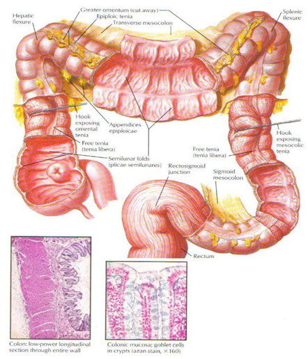
Colon
- It is also called as the large bowel or large intestine.
It has the following parts:
- Caecum
- Ascending colon (20 cm)
- Hepatic flexure
- Transverse colon (45 cm)
- Spleenic flexure
- Descending colon (30 cm)
- Sigmoid colon (35 cm)
- Rectum (12 cm)
- Anal canal (4cm)
- Taenia coli
- haustrations
Portal Circulation
Liver (Portal Triads)
- Lies in the inferior surface of liver in gall bladder fossa.
- It stores and concentrates bile.
- Dimension- 7-10 X 3 cm
- Capacity- 60 ml
Parts:
- Fundus
- Body
- Neck with hartman’s pouch
Cystic duct and CBD
- It is 4 cm duct.
- It drains into the common bile duct.
- The mucosa of the cystic duct forms 15-20 spiral folding, they are called valves of Heuster.
- CBD is 7-8 cm long and is 6 mm in diameter.
- It begins in the inferior surface of the liver by union of two hepatic ducts.
- When the cystic duct mixes with the hepatic duct, its called as CBD.
- It drains into the 2nd part of duodenum with the pancreatic duct.
- Before the opening, the distal part of this duct is dilated, that is called as the ampulla of vator, and it has a valve, called as the sphincter of ODDI.
Pancreas:
- Pan- all, creas- flesh.
- Size- 15-20 cm x 3 cm
- Lies transversly in the abdomen, between the duodenum and the spleen.
- Seperated from the stomach by lesser sac.
- It has head, uncinate process, neck, body and tail.
- The head is related with the duodenum, lies in the vicinity of the duodenum and hence its called as the romance of abdomen.
- It has two ducts- that drains the pancreatic fluid in the duodenum
- Duct of Wirsung /major duct- it drains the neck, body and tail, 3mm in diameter and drains into the CBD.
- Duct of Santorini- drains the head and the uncinate process, and open in the duodenum as separate opening.
- Head
- Uncinate process
- Body and
- Tail.
Blood supply
- Head and uncinate process- by the superior and inferior pancreaticoduodenal artery.
- The body and tail by the branches of the splenic artery.
Function
- Exocrine function
- Endocrine function
Histology:
- Acini
- Pancreatic islets (islets of langerhans)
Omentum
- The folds of peritoneum between the stomach and other visceral organs.
- It covers the coils of the intestine.
They are two,
- Greater omentum
- Lesser omentum.
Greater omentum:
- It is called as the policeman of the abdomen.
- Between the stomach and the tranverse colon.
- Has four folds of peritoneum.
- The space between the 2nd and the 3rd layer makes a sac called as lesser sac behind the stomach and the pancreas, also called as the omental bursa.
Contents:
- Right and the left gastroepiploic vessels
- Fat.
Lesser omentum:
- It is the fold of peritoneum between the lesser curvature of the stomach and the 1st part of the duodenum to the liver.
It is divided into
- Hepatogastric ligament
- Hepatoduodenal ligament
Contents:
- Hepatic artery
- Portal vein
- Bile duct
- Lymphatics
- Right and the left gastric vessels.
Mesentery
- The fold of the peritoneum that suspends the coils of intestines with the posterior body wall.
- It is fan shaped, with small root.
- The root is 15 cm and attached to the posterior body wall that runs obliquely from the left of the L2 vertebra, to the right sacroiliac joint.
- The intestinal boarder is around 6 m long.
Contents:
- Jejunal and ileal branches of the SMA
- Veins
- Nerves
- Lacteals and lymphatics
- Fat
- Transverse mesocolon, mesoappendix and sigmoid mesocolons are similar folds of the peritoneum that suspends the transverse colon, appendix and the sigmoid colon
Relations of the stomach
Anteriorly:- Liver (right part)
- Diaphragm (left upper part)
- Anterior abdominal wall (left lower part)
Posteriorly separated by peritoneum of lesser sac from the following (“stomach-bed”)
- Pancreas
- Left suprarenal gland
- Left kidney
- Spleen
- Transverse colon and transverse mesocolon
Arteries of stomach
Left and right gastric arteries - Arise from celiac trunk and proper hepatic artery, respectively.
- These two vessels run in lesser omentum along lesser curvature , and anastomose end-to-end.
Right and left gastroepiploic arteries
- Arise from the gastroduodenal and splenic artery, respectively.
- These two vessels pass into the greater omentum, run parallel to the greater curvature, and anastomose end-to-end.
Short gastric arteries
- Branches of splenic artery
- Course through the gastrosplenic ligament
- Supply the fundus of stomach.
Posterior gastric artery (72%)
- Arise from the splenic artery
- Course through the gastrophrenic ligament and supply the posterior wall of fundus of stomach.
- Right and left gastric veins empty directly into hepatic portal vein.
- Left gastroepiploic and short gastric veins drain into hepatic portal vein via the splenic vein.
- Right gastroepiploic vein drain into superior mesenteric vein.
Lymph drainage of stomach
- Right and left gastric ln. lie along the same vessels and finally to the celiac ln.
- Right and left gastroomental ln. lie along the same vessels, the former drain into subpyloric ln., the latter drain into splenic ln.
- Suprapyloric and subpyloric ln. receive lymphatics from pyloric part and finally to the celiac ln.
- Splenic ln. receive lymphatics from fundus and left third of stomach, and finally to the celiac ln.
Parasympathetic innervation
- The anterior vagal trunk divides into anterior gastric and hepatic branches
- The posterior vagal trunk divides into posterior gastric and celiac branches
- The anterior and posterior gastric branches descend on the anterior and posterior surfaces of the stomach as a rule about 1 to 2 cm from the lesser curvature and parallel to it in the lesser omentum as far as the pyloric antrum to fan out into branches called “crow’s foot” to supply the pyloric part
Sympathetic innervation
- Mainly from celiac ganglia
- Afferent and efferent fibers derives from thoracic segments (T5 -L1)
Relations of duodenum
Anteriorly
- Quadrate lobe of live
- Gallbladder
Posteriorly
- Common bile duct
- Gastroduodenal a.
- Hepatic portal v.
- Inferior vena cava
Superioely
- Omental foramen
Inferiorly
- Head of pancreas
Relations of descending part (2nd)
Anteriorly
- Liver
- Transverse colon and transverse mesocolon
- Loops of small intestine
Posteriorly
- Right renal hilum and ureter
- Right renal vessels
Medially
- Head of pancreas
- Common bile duct and pancreatic duct
Laterally
- Right colic flexure
Relations of horizontal part (3rd)
Superiorly
- Head of pancreas
Inferiorly
- Loops of small intestine
Anteriorly
- Radix of mesentery
- Superior mesenteric a. and v.
Posteriorly
- Right ureter
- Inferior vena cava
- Abdominal aorta
Relations of ascending part (4th)
- Right — Head of pancreas and abdominal aorta
- Left — left kidney and ureter
- Arteries Superior pancreaticoduodenal a. (branch of hepatic artery….coeliac trunk)
- Inferior pancreaticoduodenal a. (branch of superior mesenteric artery)
- Spleenic artery.
- Veins follow arteries, draining directly into superior mesenteric and hepatic portal veins
Relations of liver
separated by diaphragm from the following
Right costodiaphramatic recess and lung
Cardiac base
Visceral surface
Left lobe is related to the stomach and abdominal part of esophagus
Right lobe is related to the right colic flexure anterioly, gallbladder and superior duodenal flexure medially, right kidney, superarenal gland posteriorly
Divisions and relations of common bile duct
Divisions Supraduodenal segment
Retroduodenal segment
Pancreatic segment
Intraduodenal segment
Supraduodenal segment
- Descends along the right margin of hepatoduodenal lig.
- To the right of proper hepatic a.
- Anterior to hepatic portal v.
Retroduodenal segment
- Behind the superior part of duodenum
- Anterior to the vena cava
- To the right of the hepatic portal v.
Pancreatic segment
- Lies in a groove between posterior surface of head of pancreas and duodenum
Intraduodenal segment
- Enters the wall of descending part of duodenum obliquely where jions the pancreatic duct to form the hepatopancreatic ampulla
- Opens at the major duodenal papilla
Cystohepatic triangle (Calot’s Triangle)
Boundaries
- Common hepatic duct on the left
- Cystic duct on the right
- Liver superiorly
Content: cystic artery, lymphatics and fat.
The Pancreas
Four parts- Head -Lies within the cancavity of the C-shaped curvatune of duodenum
- Uncinate process- a projection to the left from the lower part of the head behind the superior mesenteric vessels.
- Neck-narrow part, overlies the superior mesenteric vessels and beginning of the portal vein
- Body-triangular in cross section, passes upward and to the left across the midline
- Tail-extends to the hilum of spleen in the splenorenal ligament
Relations of pancreas
Head of pancreas
- Located in C-shapes curvature of doudenum
Anteriorly
- Transverse mesocolon
Posteriorly
- Inferior vena cava
- Right renal vessels
- Common bile duct
Neck of pancreas
- Anteriorly-pylorus
- Posteriorly-beginning of hepatic portal v.
Body of pancreas
Anteriorly
- Separated from stomach by omental bursa
Posteriorly
- Abdominal aorta
- Left suprarenal gland
- Left kidney
- Left renal vessels
- Spleen vein
Superiorly
- Celiac trunk
- Celiac plexus
- Splenic a.
Relations of spleen
Visceral surface
Anteriorly-fundus of stomach
Posteriorly-left suprarenal gland and kidney
Inferiorly-tail of pancreas and left colic flexure
Position-it lies between the parietal peritoneum and transvers fascia of the posterior abdominal wall, from diaphragm to promontory of sacrum, continuation with extraperitoneal fascia.
Contents
- Kidney
- Suprarenal gland
- Ureter
- Abdominal aorta
- Inferior vena cava
- Nerves and lymphatics
- Loose connective tissue
Kidney
- Paired structures in the retroperitoneum.
- Size 11x6x3 cm.
- The right kidney is slightly lower than the right kidney due to relative position of the liver.
- The kidney lie in the lumbar region, but it occupies some space in the hypochondrium, and umbilical regions.
- Each kidney has two parts, the medulla and the cortex.
- Microscopically, each kidney has its functional units that are called as nephrons.
Right kidney
Anterior-
- liver,
- 2nd part of diaphragm,
- hepatic flexure and small intestines.
- Adrenal gland
- Ureter
- Diaphragm
- Muscles- psoas, quadratus lumborum, tranversus abdominis.
- Subcoastal illiohypogastric and illioinguinal nerves
- Rib 12th.
Antrior:
- Adrenal
- Spleen
- Stomach
- Pancreas
- Spleenic flexure
- Jejunum
- Adrenal gland
- Ureter
- Diaphragm
- Muscles- psoas, quadratus lumborum, tranversus abdominis.
- Subcoastal illiohypogastric and illioinguinal nerves
- Ribs 11th and 12th.
- Fibrous capsule
- Perirenal fat
- Gerota’s fascia.
- Artery- renal artery, 30 % will have accessory renal artery.
- Vein- renal vein
- Lymphatic drainage
- To paraaortic lymph nodes.
Shape and position
- Right is pyramidal in shape, left one semilunar in shape, consisting of out cortex and inner medulla
- Located retroperitoneally, superomedial to superior poles of each kidney, enclose with the kidney by the renal fascia
Arteries
- Superior suprarenal a.
- Middle suprarenal a.
- Inferior suprarenal a.
- Right suprarenal v. drains into inferior vena cava
- Left suprarenal v. joins left renal v.
No comments:
Post a Comment